This tutorial will take you through the basic structure and function of the back part of the eye, where you detect light and send information about it to your brain.
You can navigate through this tutorial using the buttons at the top of the screen.
The tutorial will ask you questions. Click on your chosen answer to see feedback; click the answer again to make the feedback disappear. When you're finished with one page, click the navigation button for the next page to move ahead.
Have fun! Click on button '1' to see the first page of the tutorial.
Page 1
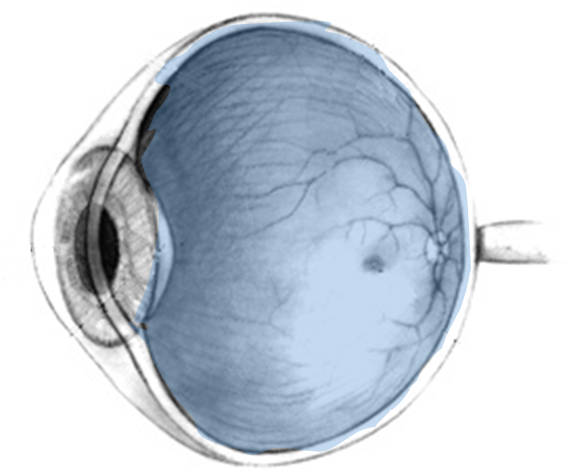
Image modified from Wikipedia. Used under a Creative Commons license
The part of your eye behind the iris and lens is the:
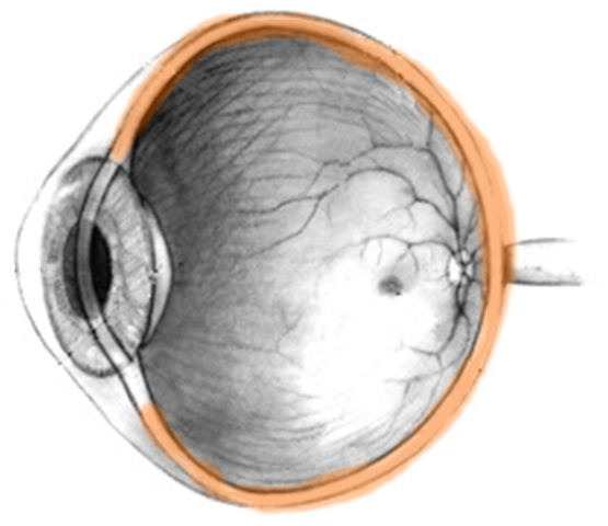
Image modified from Wikipedia. Used under a Creative Commons license
The white layer covering the outside of the eye is the:
Light rays are detected when they hit the inner layer, the:
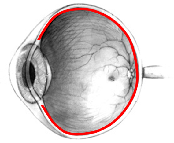
Image modified from Wikipedia. Used under a Creative Commons license
The layer of blood vessels between the sclera and retina is the ________
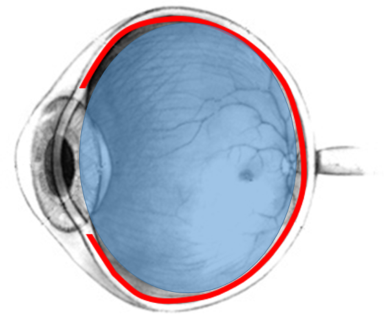
Image modified from Wikipedia. Used under a Creative Commons license.
The jelly-like substance that fills the posterior chamber, pushing the retina against the choroid, is the _______
Page 2
Good work on the anatomy!
The posterior chamber of your eye is all about REPORTING light.
The photoreceptors in the retina change their firing in response to light, and transmit that information to the brain.
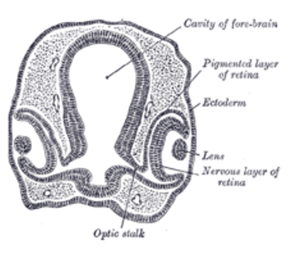
Image from Wikipedia. Used under a Creative Commons license.
In this section through an embryonic brain, you can see the hollow brain in the center and the retinas developing as outpouchings on each side of it. They are actually brain tissue! The skin over each developing retina will turn into the cornea and anterior chamber of that eye.
This is the brain and retinas of an early chick embryo, but humans develop the same way. So your retinal cells are nervous cells, like the nerve cells in your brain. They are specialized, however, to respond to light.
Page 3
The light-detecting nerve cells in your eyes are called photoreceptors.
Let's have a look at them up close:
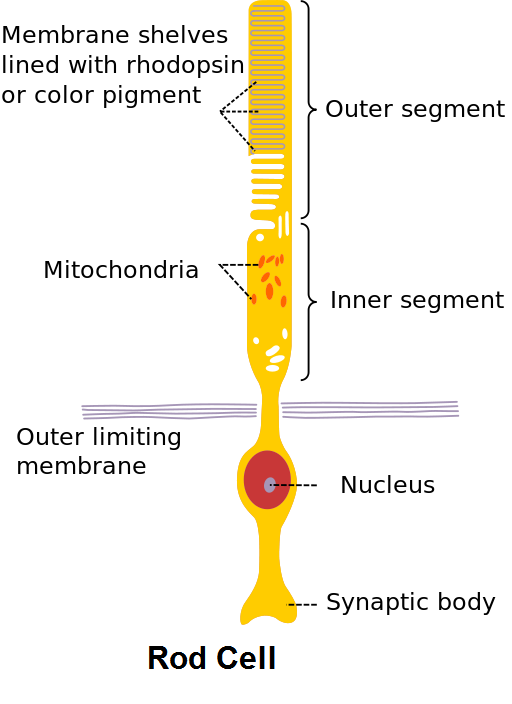
Modified from image by Madhero88, from Wikipedia. Used under a Creative Commons license.
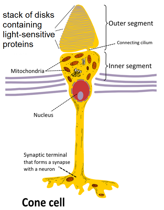
Modified from image by Ivo Kruusamägi (2010), from Wikipedia. Used under a Creative Commons license.
Each one has a cell body with a nucleus, just like any neuron. Below the cell body you see an axon stretching down to a synapse, where it will pass information to another cell. Again, this is just what you expect to see in a neuron.
It's up above the cell body that these photoreceptors look a bit different. Instead of hair-like dendrites, each of them has an expanded area full of mitochondria. These cells must use a lot of energy! And above that is an area full of folds in the membrane. It looks a little like a stack of pancakes inside the cell - and on these pancakes are the pigmented proteins that serve as receptors to catch the light.
To understand how photoreceptors fire, you need to first remind yourself of the second messenger system. Remember, your cells use this system when they are receiving a stimulus at one place on the cell - but the reaction needs to happen at another place, far away from the stimulus.
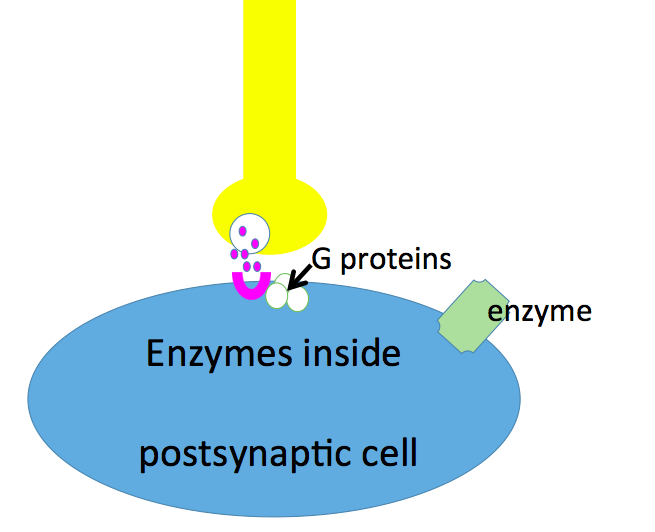
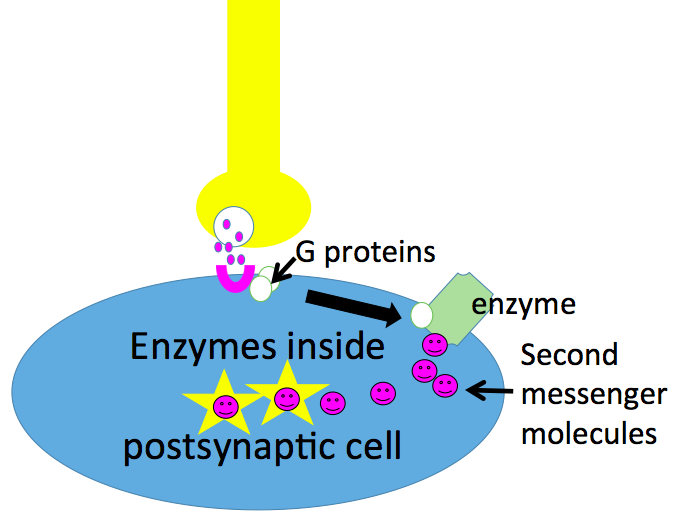
In this series of cartoons, you see a neuron stimulating a cell. The neuron is releasing neurotransmitters that attach to receptors on that cell's surface - but the desired response is going to happen way down inside the cell, where the neurotransmitters and receptors can't go.
In the second frame of the cartoon, then, you see that the receptor that has received the neurotransmitter is sending a small protein called the G protein across the cell's surface to an enzyme. The G-protein affects the enzyme's activity, causing it to produce small second messenger molecules that can diffuse down inside the cell and make enzymes deep inside respond.
An analogy for this is a light switch on the wall of your room. The switch is the receptor. When your finger hits it, it sends a message along the wires to the light fixture in the ceiling; the message is like the G-proteins, running across the cell's membrane to the enzyme. Just like the enzyme on your cell membrane, the light fixture in the ceiling produces something that can go down inside the room and affect things that are nowhere near the switch, or the wire, or the light bulb; it can start you studying.

Modified from "Woman studying cartoon", Public Domain Pictures
Page 4
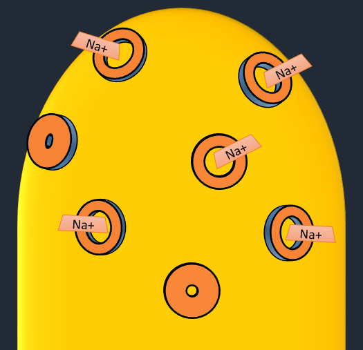
Here's the top of a photoreceptor, in the dark. Like all nerve cells, this one has Na+ channels that can open or close - and right now, they are open. What is happening to the cell as a result?
Na+ is moving into the cell and it is depolarizing
Na+ is moving into the cell and it is repolarizing
Na+ is moving out of the cell and it is depolarizing
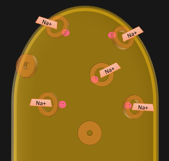
What made those Na+ channels open? Let's look inside. See the little pink second messenger molecules? Those molecules made the channels open and let the Na+ in, making the cells depolarize. So in the dark, this photoreceptor is firing.
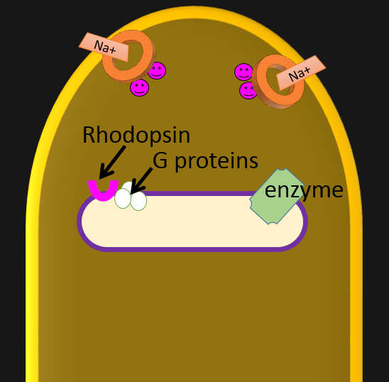
Now let's add one of those membrane 'pancakes' inside the cell. In real life there are a whole stack of them. This pancake has some important things on it, starting with rhodopsin, the protein that receives light. It's dark right now, so the rhodopsin is purple and it isn't doing anything - and neither are the G proteins attached to it, or the enzyme a little further away on the same pancake.
Up at the surface of the cell you can see some of those open Na+ channels and the little second messengers that made them open.
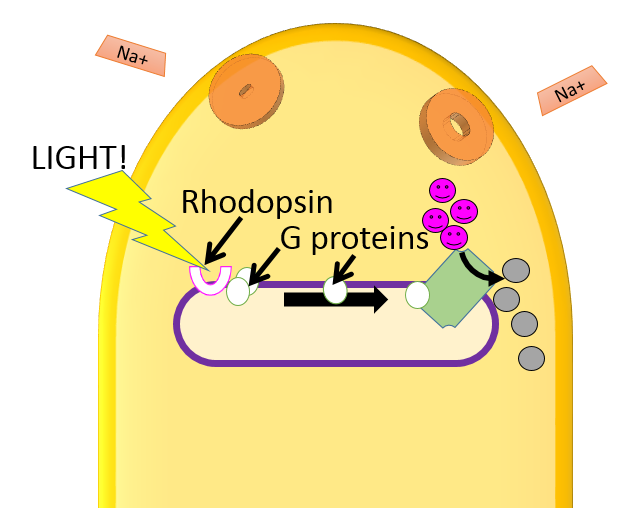
Add light and everything changes!
The light hits the rhodopsin and makes it change shape - and color. It bleaches.
Then the G-protein reacts, and one of its units moves over to the enzyme.
Then the enzyme is activated, and starts doing its job - to turn those little second messengers into an inactive form.
When the second messengers are taken away, the Na+ channels close.
When the channels close, Na+ stops entering the cell and the cell stops firing...
and that's what sends a message to your brain that some light has hit that cell.
Page 5
Time for a review! See if you can fill in the blanks in this flow chart of how a photoreceptor fires.
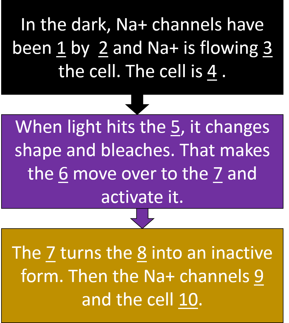
When light hits the (5) membrane / ion channels / rhodopsin, it changes shape and bleaches. That makes (6) G proteins / receptors / neurotransmitters
move over to the (7) ion channels / receptors / enzyme and activate it.The (7) turns the second messengers / ion channels / G proteins into an inactive form. Then the Na+ channels (9) close / open / and the cell (10) fires / stops firing .
Page 6
Good work summarizing the events!
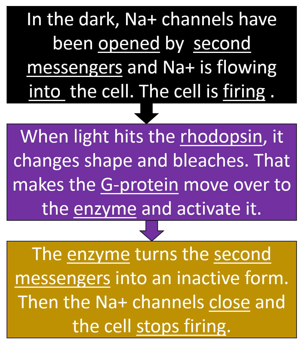
Now let's zoom out and look at the retina, the layer made of thousands of these photoreceptors lying side by side - and the nerve cells that are attached to them.
Page 7
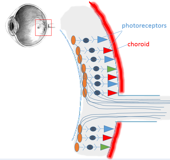
Image modified from Wikipedia. Used under a Creative Commons license
Let's zoom out a little and see how those cells are arranged in the back of your eye.
The photoreceptors are in the posterior side of the retina, right next to the choroid blood vessels so they can get lots of nutrients and Oxygen.
The photoreceptors in this picture are named for their shape. I drew the photoreceptors in different colors to indicate that they respond to light of different colors- red, green, and blue. What's their name?
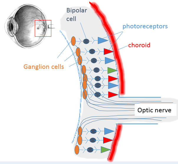
Image modified from Wikipedia. Used under a Creative Commons license
The photoreceptors synapse with the bipolar cells.
The bipolar cells synapse with the ganglion cells.
The ganglion cells send their axons out through the optic nerve, all the way into the brain.
Page 8
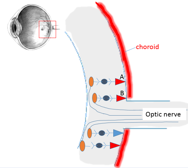
Image modified from Wikipedia. Used under a Creative Commons license
Now I’ll take some of them away so you can see more clearly.
Suppose you look at two objects. The light from one focuses and hits cone A, and the light from the other hits cone B.
Can your brain tell which cone the light hit?
No, because they both detect red light
No, because they’re both cones
Yes, because they have separate connections to the brain
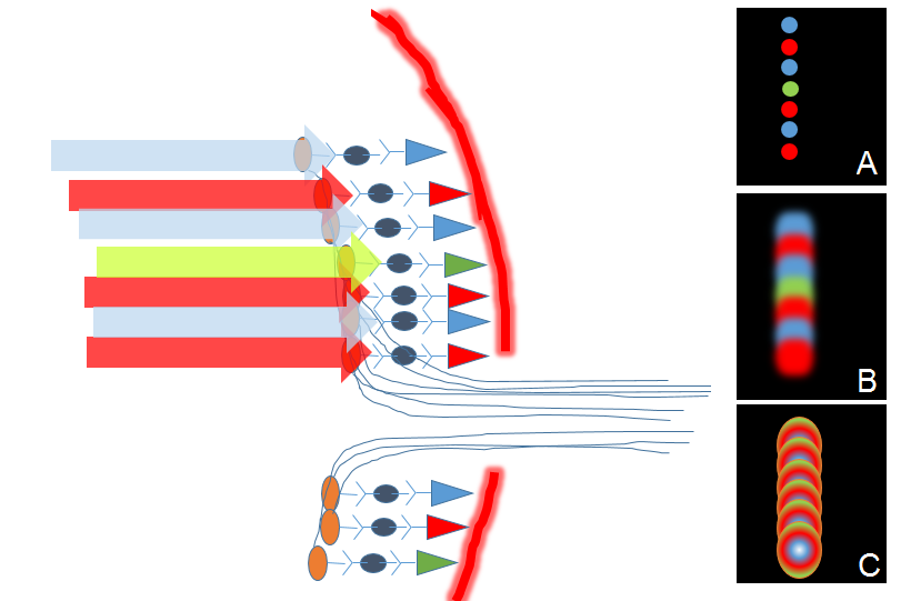
Image modified from Wikipedia. Used under a Creative Commons license
If light hits all these cones at once, what will your brain detect?
Each of your cones has its very own bipolar cell and ganglion cell – its own hotline to the brain!So your brain can tell what each individual cone is seeing. That’s how you can distinguish small objects from one another.

So cones give you acute color vision. The down side is – they need a lot of light to work. They are not very SENSITIVE in low light.
Page 9
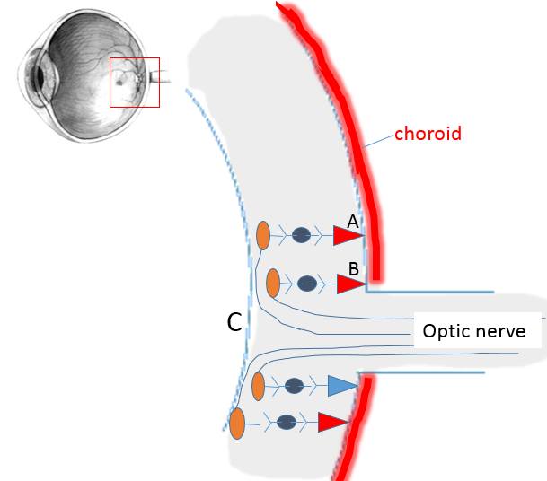
Image modified from Wikipedia. Used under a Creative Commons license
Before we go away from this diagram of the back of your retina, answer another question.
What will happen if light focuses on point C of this retina?
A big blur of light, because all the axons are stimulated
Nothing, because it won’t hit any photo-receptors
White light, because it won’t hit any cones
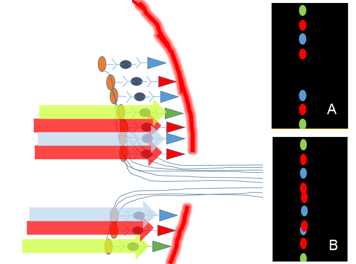
Image modified from Wikipedia. Used under a Creative Commons license
Where the axons exit your retina into the optic nerve, you have a BLIND SPOT – a place where there are no photoreceptors to detect light.
But in real life, what does it look like to you when light hits this spot?
Page 10
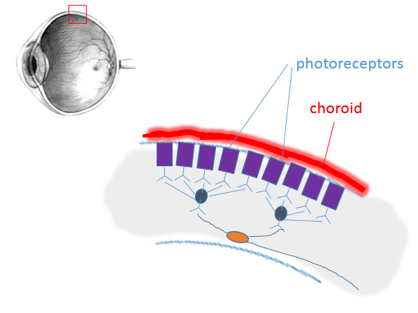
Image modified from Wikipedia. Used under a Creative Commons license
Let’s look at a close-up of a different part of the retina – toward the edge.
Just like before, the photoreceptors are right next to the choroid blood vessels so they can get lots of nutrients and Oxygen. But they look different.
What do you think these photoreceptors are called?
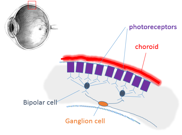
Image modified from Wikipedia. Used under a Creative Commons license
Just like cones, rods synapse with bipolar cells. The bipolar cells then synapse with ganglion cells.
But it’s not quite the same, is it? What's different?
There aren’t enough bipolar cells
There aren’t enough ganglion cells
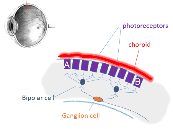
Image modified from Wikipedia. Used under a Creative Commons license
If light hits cell A on this part of the retina, can your brain tell it apart from light hitting cell B?
No, they use the same axon to the brain
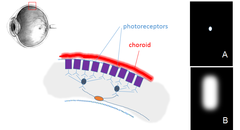
Image modified from Wikipedia. Used under a Creative Commons license
Your rods provide sensitive vision in low light - but it is not in color, and it is not ACUTE. You can't distinguish things well using your rods.
Page 11
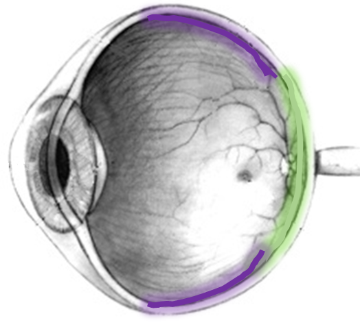
Image modified from Wikipedia. Used under a Creative Commons license
The cones (colored green in the drawing) are located in the back of your eye, in the central portion of your retina. The rods (colored purple in the drawing) are located in the peripheral parts of your retina.
That means cones see what’s right in front of you, while rods see the things that are off to the sides.

Your eyes actually send a message like this to your brain.

Image modified from Wikipedia. Used under a Creative Commons license
How do you see the whole scene in color? You keep moving your eyes back and forth, and your brain remembers the colors you just looked at. It fills them in, the same way it fills in your blind spot.
Page 12
In this figure, you’re looking down on someone’s eyes and brain. The two sides of the retina and their nerve tracts have been color-coded; the left side of each retina is orange and the right side is blue.
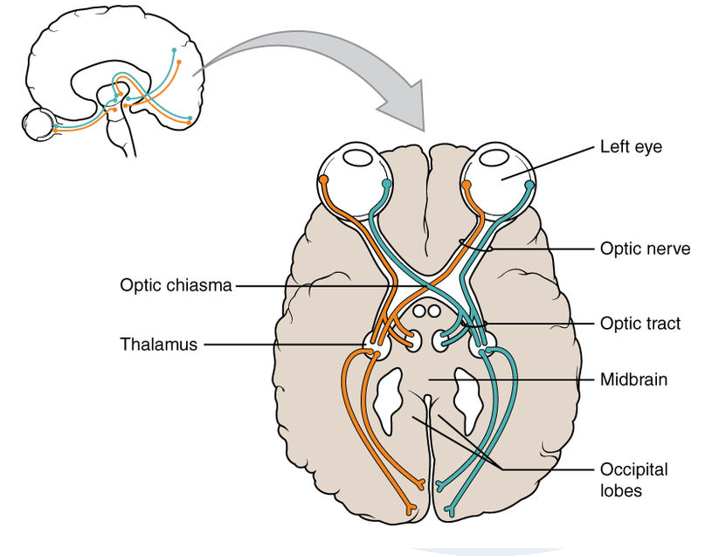
Image modified from Wikimedia Commons. Used under a Creative Commons license
Which cells in the retina would see things in the left end of the title bar?
Which cells in the retina would see things in the right end of the title bar?
Page 13

Image modified from Wikimedia Commons. Used under a Creative Commons license
You figured out that light from something at the left would fall on photoreceptors on the RIGHT side of the retina - the blue photoreceptors.
Which side of the BRAIN will get the message?
the right side
the left side
both sides
Click here for a summary:
Page 14
What’s the point of this? Why should fibers from your eye cross over to the other side of the brain?
Here’s a clue: it only happens in animals with binocular vision – animals that can look at one object with both eyes at once.
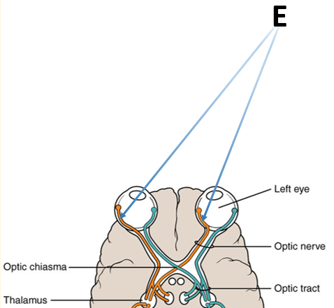
Image modified from Wikimedia Commons. Used under a Creative Commons license
When both eyes look at the ‘E’ up on the right, the light hits the left side of the retina in both eyes. The nerve impulses caused by that image are sent to the same place in the left side of the brain. The brain can compare them.
Look at where those light rays hit the retina – almost the same place in each eye. That means the nerve impulses sent from the retina will end up in almost exactly the same place in your brain. Your brain will know that you saw that object, and that the images from your two eyes were very similar.
Now, let's look at an E much closer to your eyes.
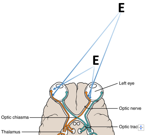
Image modified from Wikimedia Commons. Used under a Creative Commons license
Where does the light hit the retinas now?
It’s a quite different spot on each retina, isn’t it?
When these messages reach the brain, they won't be that close together. The brain will see that the images from your two eyes are quite different from each other. That tells your brain the object is closer to your eyes.
This is what gives you depth perception - and it's why you have a harder time judging distances if you close one eye. Your brain tells distance by comparing how something looks to your right eye with how it looks to your left eye. The further away the object is, the more alike it will look to your two eyes.
Page 15
One last thing to notice in this diagram is that as each optic tract enters the brain, it sends a little branch in toward the center of the brain. These branches end in structures called the superior colliculi. This area controls reflex responses to sight. So you sometimes react right away, even before your brain knows what you saw!
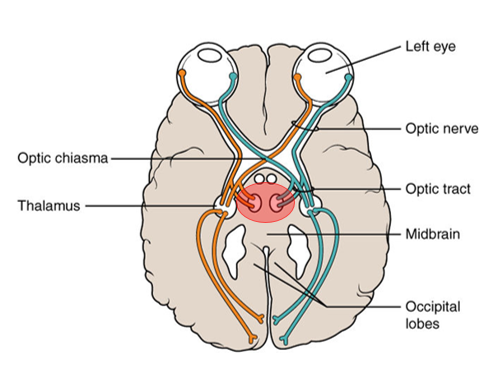
Image modified from Wikimedia Commons.
Now you should be comfortable with rods, cones, and how the visual impulse reaches your brain.
Happy studying!
This tutorial is based on material from Fox, S.I., 2013. Human Physiology, 13th ed. McGraw-Hill.