Introduction
This tutorial will take you through the basics of skeletal muscle contraction and some examples of how it can be altered.
You can navigate through this tutorial using the buttons at the top of the screen.
The tutorial will ask you questions. Click on your chosen answer to see feedback; click the answer again to make the feedback disappear. When you're finished with one page, click the navigation button for the next page to move ahead.
Have fun! Click on button '1' to see the first page of the tutorial.
Page 1
To begin with, let's review how the nervous system controls the muscles.
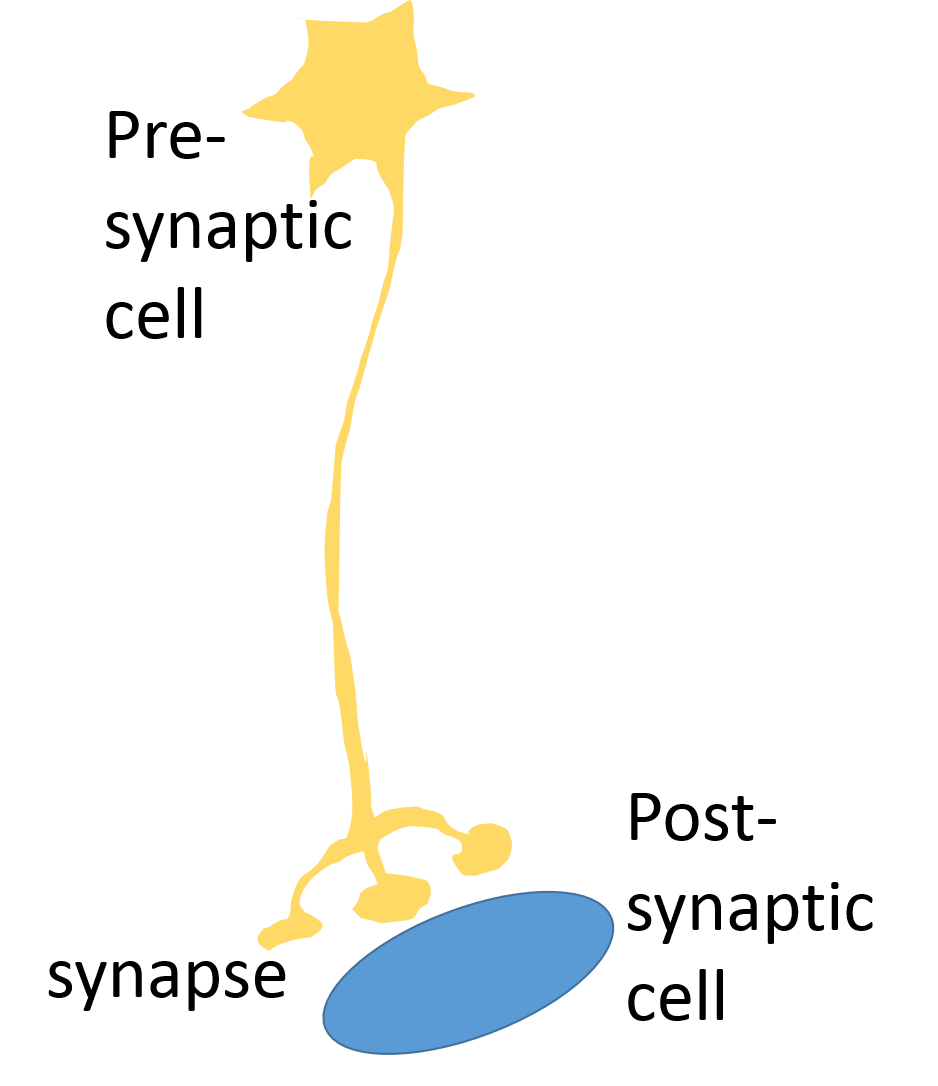
Here's a presynaptic motor neuron, sending its axon down to a postsynaptic muscle. The axon branches at its lower end, and its branches connect with a bunch of muscle cells inside this muscle. The muscle cells controlled by one motor neuron are called its motor unit.
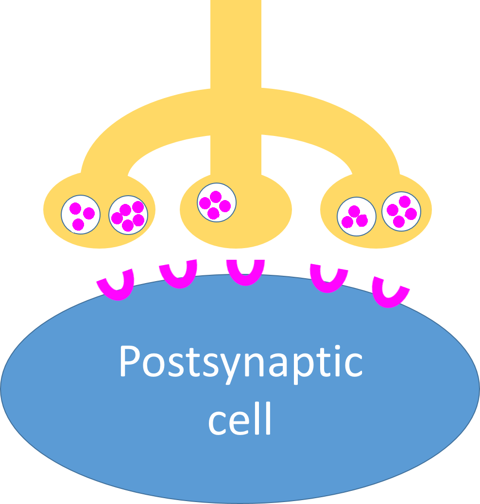
Here's a closer look at the neuromuscular synapse. The presynaptic neuron contains vacuoles full of the neurotransmitter acetylcholine; the postsynaptic cell has nicotinic acetylcholine receptors on its membrane. This area where the nerve cell meets the muscle is called the motor end plate.
Page 2
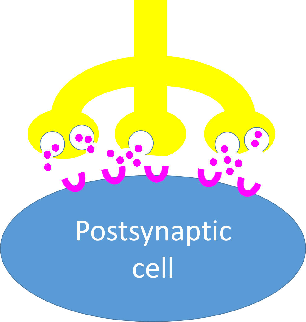
When the motor nerve fires, it will release the acetylcholine into the synapse.
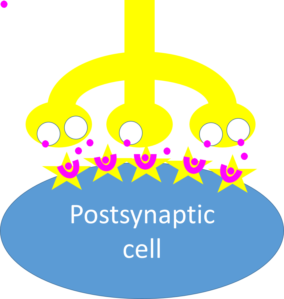
The acetylcholine diffuses across the synapse and attaches to the nicotinic receptors, causing ion channels to open in the muscle cell's membrane. Now Na+ can diffuse into the muscle cell, and it too will fire - but only as long as the acetylcholine is on the receptors.
Soon, the acetylcholine will be removed by the enzyme acetylcholinesterase and the muscle firing will stop.
What would happen to muscle firing if the acetylcholinesterase didn't work?
What if the presynaptic neuron wouldn't release acetylcholine?
What if the nicotinic receptors were blocked so acetylcholine couldn't attach?
Page 3
Now let's have a look at this muscle cell that is being stimulated. It doesn't look at all like the blue blob in the pictures so far.Muscle cells are long and thin, which is why they're called myofibers. They have many nuclei, which makes sense; the nuclei contain instructions for creating proteins, and muscle cells are full of protein.
The parts of muscle cells have different names from ordinary cells: their cytoplasm is called sarcoplasm, and their cell membrane is called the sarcolemma.
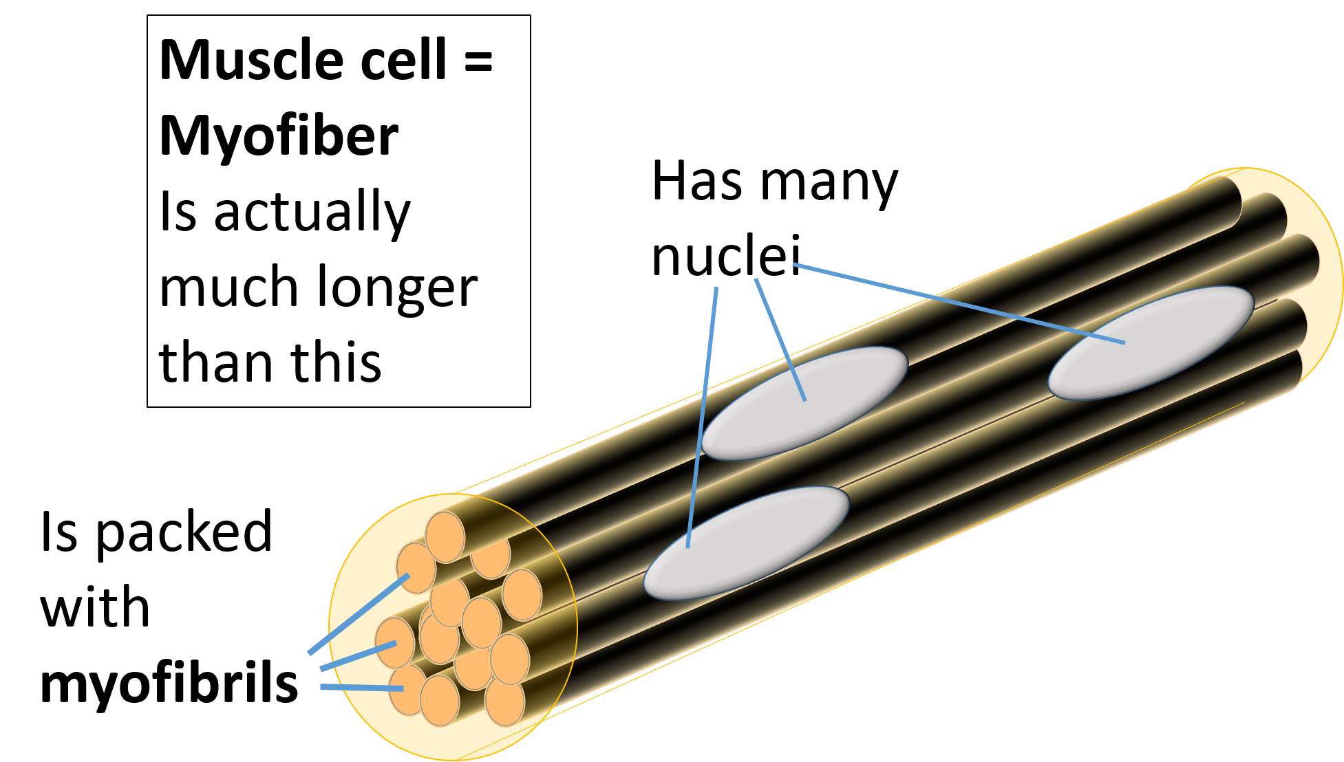

Each myofibril is divided into segments called sarcomeres. The walls between the sarcomeres are called Z-disks.
So your muscles can pull, but they cannot push.
Page 4
Each of the myofibrils is connected to the external membrane (sarcolemma) of the myofiber by many transverse tubules or T- tubules that reach in and wrap around the myofiber.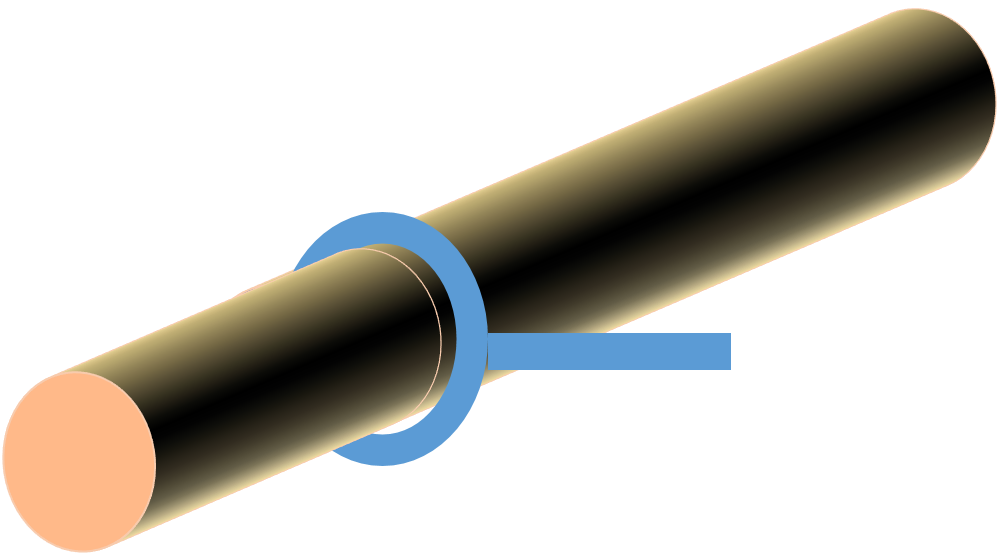
On each side of the T-tubules, a network of hollow membranes lies on the surface of the myofibril.
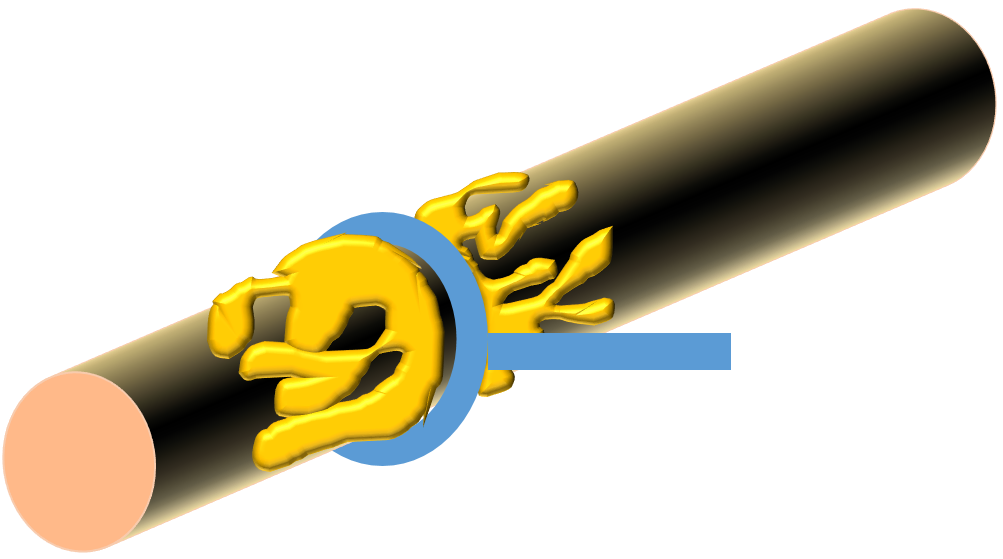
The sarcoplasmic reticulum is full of Ca2+ ions.
Page 5
We've seen what lies on the surface of each myofibril -- the T-tubule and the sarcoplasmic reticulum -- but what's inside?Each myofibril is full of tinier fibers -- protein fibers. These are the contractile proteins, that can actually make the myofibril contract. But they are packed into the inside of the myofibrils, and the T-tubule has only carried the electric impulse to the surface of the myofibrils.
You've seen something like this before. When something makes the surface of a cell fire, but the impulse activates something down inside the cell, it's usually because some molecule, a second messenger, was released and diffused down inside to the cell to activate proteins.
Ca2+ can act as a second messenger, and the sarcoplasmic reticulum is full of Ca2+.
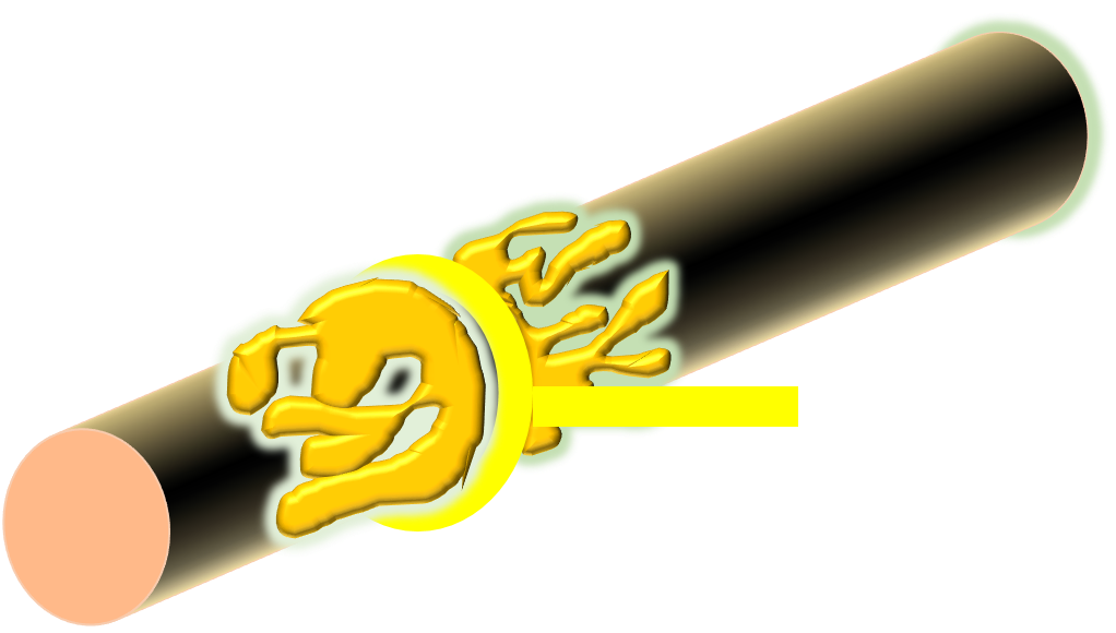
So when the T-tubules stimulate the sarcoplasmic reticulum, it will release the Ca2+ and the Ca2+ will diffuse down inside the myofibrils and activate the contractile proteins.
Page 6

1. Remember the sarcomeres, the segments each myofibril was divided into? The walls between the sarcomeres are called Z-disks. Each sarcomere contains two contractile proteins - actin and myosin.

2. Filaments of the protein actin are atttached to the z-disks. They extend out into the sarcomere. So there are actin filaments extending into the sarcomere from each end - from each z-disk.

3. Filaments of another protein, myosin, lie in the center of the sarcomere extending toward each wall until their ends overlap the actin filaments.

4. When the sarcomere contracts, the actin filaments slide past the myosin filaments, toward the center of the cell, pulling the z-disks they are attached to together.. The actin and myosin filaments don't get any shorter, they just slide past each other.
As a result, the sarcomere gets shorter. When every sarcomere gets shorter, the myofilament will too - and when every myofilament gets shorter, the whole muscle will contract.
Page 7
What made the filaments slide past each other? Let's look closer.

Here's a diagram of one sarcomere, showing the actin filaments extending inward from the z-disks and the myosin filaments lying in the center of the sarcomere. See the crossbridges coming out from the myosins and grabbing the actins? Those crossbridges can move, pulling the actins toward the center of the sarcomere. That's what makes it get shorter.
Moving the crossbridge uses ATP. That's why your muscles need so much energy. And after the myosin has pulled, when it lets go of the actin - that needs ATP too! You can't either contract or relax your muscles without ATP.
What would happen to a person whose muscles had no ATP at all?
Page 8
So myosins can pull actins if they have ATP.
As long as you're alive, your muscles have ATP - so why aren't they always contracting?
The answer is that there's something in the way, keeping the myosins from grabbing the actins.
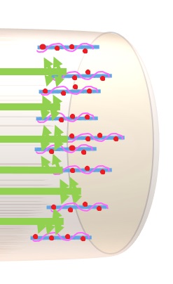
See the pink proteins wrapped around the actin? Those are tropomyosin. The red dots are troponin proteins. They are covering up the binding sites where myosin can grab actin - so as long as they are in position, the muscle won't be able to contract.
Actin and myosin are called contractile proteins because working together, they make the sarcomere contract. Troponin and tropomyosin are called regulatory proteins because they control whether actin and myosin can work or not.
The muscle will not be able to contract unless something moves the regulatory proteins off the binding sites.
Page 9
I bet you can guess what might move into that sarcomere, when the muscle cell has been stimulated, and make the regulatory proteins get off the binding sites.
 Remember the Ca2+ that leaked out of the sarcoplasmic reticulum when the cell fired, and diffused down into the myofibril? That's the link!
Now you should be able to put it all together into a story from start to end. Click on the right word in each sentence.
Muscle contraction starts when a motor neuron / sensory neuron fires and releases the neurotransmitter norepinephrine / acetylcholine into the neuromuscular synapse. It diffuses across the synaptic gap and attaches to muscarinic receptors / nicotinic receptors, causing the muscle to open ion channels in its sarcolemma / sarcoplasm and depolarize. The cell will keep firing until the neurotransmitter is removed by acetylcholine / acetylcholinesterase.
The muscle cell or myofiber / myofibril is packed full of myofibers / myofibrils which are divided into small contractile units called sarcolemma / sarcomeres. These are separated from one another by dividing walls called Z-disks / I-bands. Filaments of the contractile protein actin / myosin are attached to each dividing wall, and filaments of troponin / myosin lie in the center of each subunit, waiting to grab those filaments and pull them, moving the walls closer together. The only thing stopping this contraction is the regulatory proteins actin and myosin / troponin and tropomyosin, which are blocking the binding sites so the contractile proteins cannot attach to each other.
When the muscle cell fires, the impulse runs across its membrane and down tubules connected to the membrane called Transverse tubules / myofibrils. The impulse is carried deep into the cell, to every part of it. Inside the cell, the impulse stimulates the sarcoplasm / sarcoplasmic reticulum to release Ca2+ / acetylcholine into the myofibrils. It attaches to the regulatory proteins, moving them out of the way and allowing contraction to start.
Page 10
Here it is in a handy flow chart.
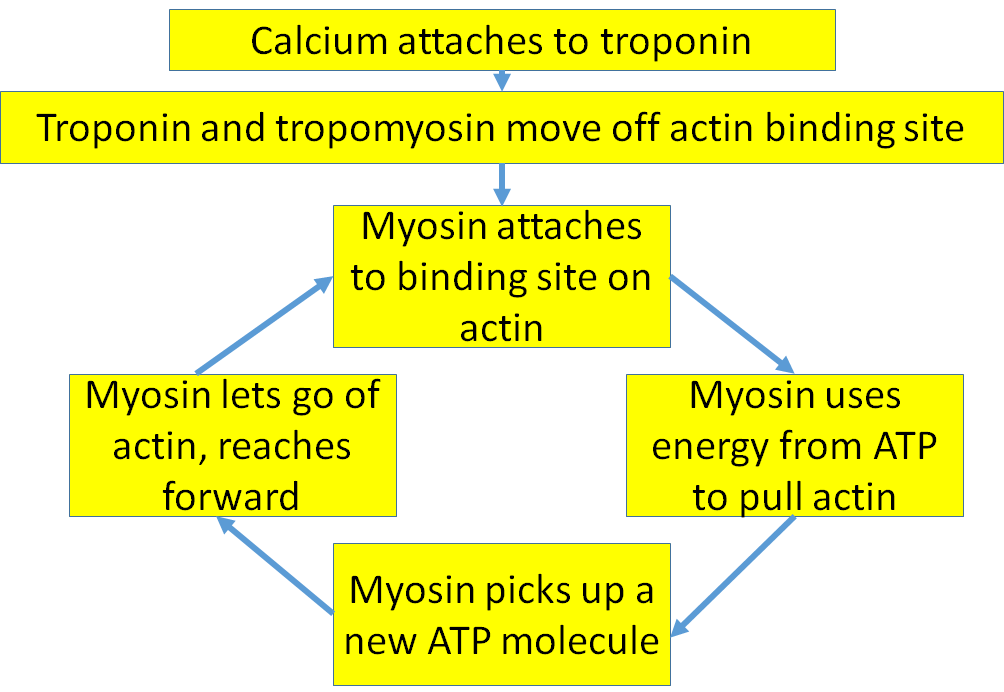
But how can this muscle ever stop contracting? The myosin just keeps grabbing and pulling.
It'll stop when it runs out of ATP
It'll stop when the actin is pulled to the middle and can't go any further.
It'll stop when the acetylcholine is removed from the receptors.
It'll stop when the Ca2+ goes away.
Sources used in creating this tutorial include:
Seeley, R.R. , Stephens, T.D., and P. Tate, 2003.Anatomy and Physiology, 6th Edition. McGraw-Hill Higher Education.