Introduction
This tutorial will take you through the structure of the ear and how it allows you to hear and keep your balance.
You will navigate through this tutorial using the buttons at the top of the screen.
The tutorial will ask you questions. Click on your chosen answer to see feedback; click the answer again to make the feedback disappear. When you're finished with one page, click the navigation button for the next page to move ahead.
Have fun! Click on button '1' to see the first page of the tutorial.
Page 1
HAIR CELLS have nothing to do with your hair. They are specialized sensory cells that detect motion.
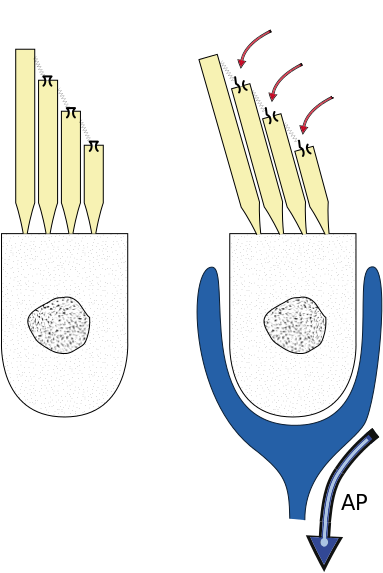
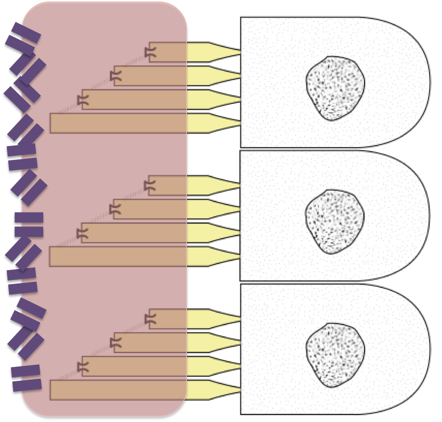
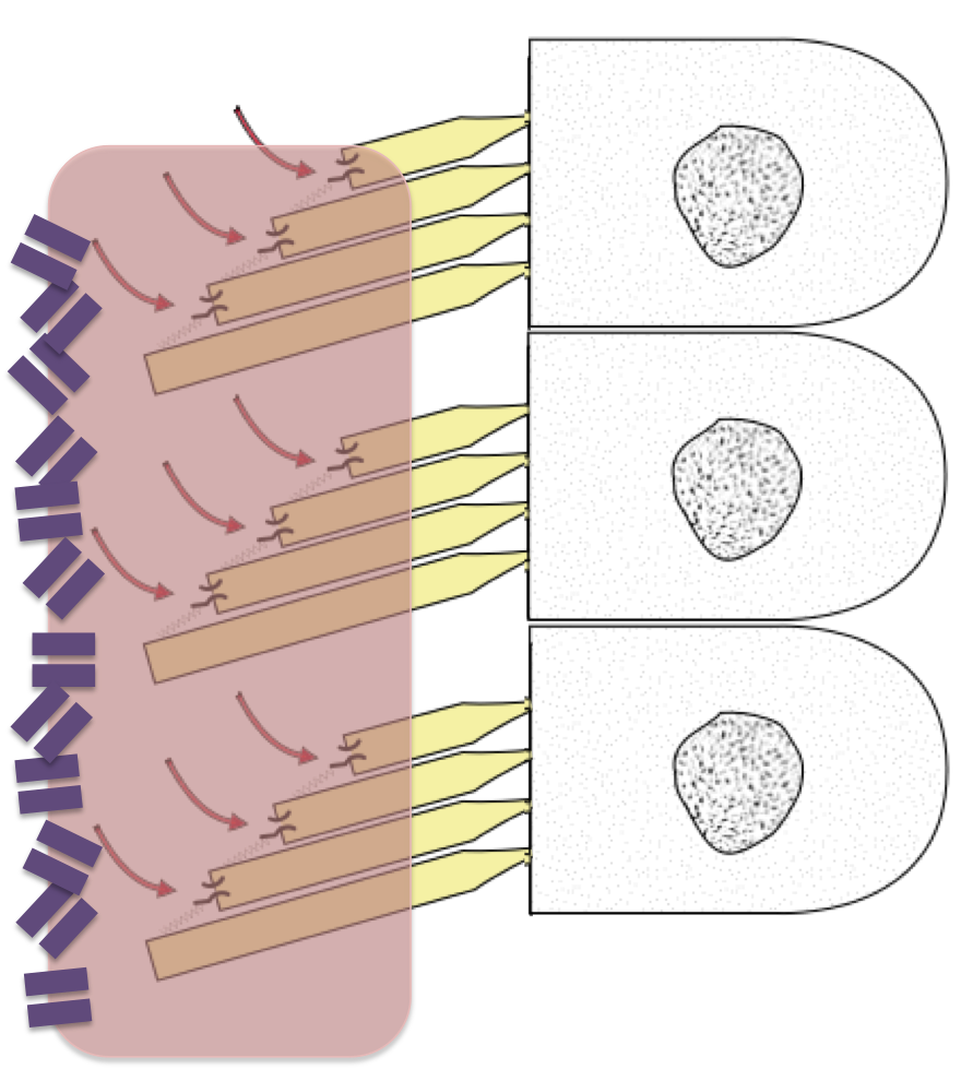
Thomas.haslwanter, 2011. HairCell Transduction, modified from Wikimedia Commons. Used under a Creative Commons license.
Hair cells have tiny hairlike cilia extending from them, and these cilia are attached to ion channels on the neighboring cilia. In the first picture, you can see the cilia sticking up from the cell and the ion channels on top of the cilia. See how the channels on neighboring cilia are attached to one another?
In the second picture the cilia have swayed to one side, and the connections between them have pulled their ion channels open. Ions can now flow into the cell and make it fire.
So anything that moves the cilia can affect the cell firing, generating an action potential that can then be sent to the brain. That's how your brain knows about the motion.
Hair cells are especially useful for detecting waves or vibrations that make the cilia sway back and forth, opening and closing the ion channels. You use them to detect sound vibrations and movements of your head; you also have some that you use to detect your head's position.
These hair cells are very delicate. They live inside chambers in your temporal bone - the chambers of your inner ear. When they're damaged, it can affect your hearing and your sense of balance.
Page 2
The structures of the inner ear protect the hair cells, but they also control what kinds of motions can affect the hair cells. The hair cells live in fluid-filled chambers, so they only detect motions in the fluid within those chambers - and those chambers are carefully designed so the fluid within them only moves in response to particular situations.
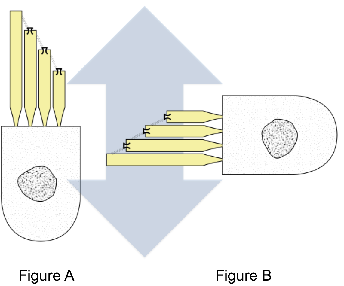
Let's look at the hair cells that tell whether you are moving up or down. These are the ones that tell you whether the elevator has started or stopped.
I bet you can figure out some of the important things about these hair cells. To start out with, how should they be oriented to detect up-and-down motion in the fluid surrounding them? Vertically, like Figure A, or horizontally, like Figure B?
Page 3
Now, let's figure out what kind of bony chamber these cells should be located in.
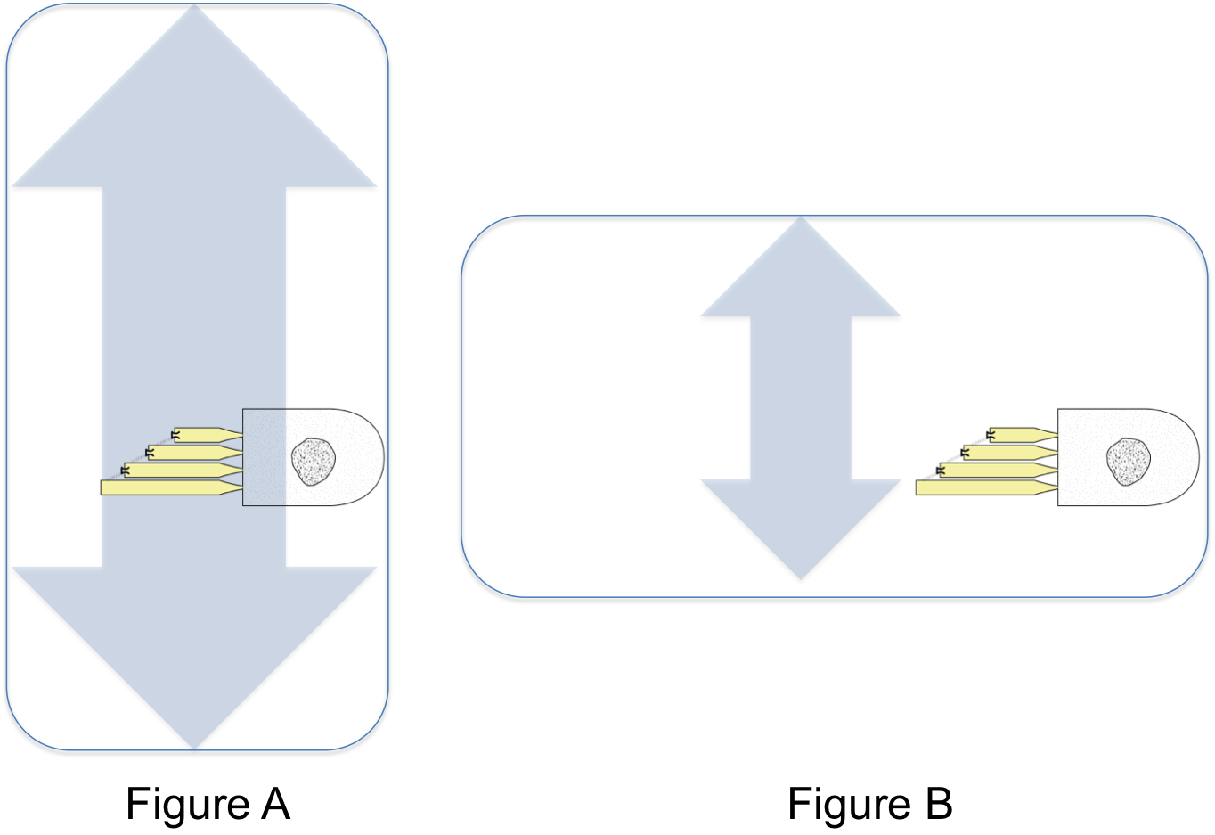
The chamber holding these cells is called the saccule. The hair cells are atteched to one side of it, their cilia sticking out into the fluid it contains. And there's one more thing to make them even more responsive to movement: a blob of jelly is stuck on the end of the cilia.
You can see the red jelly in this picture, with all the hair cells' cilia stuck into it. The jelly has a lot of tiny 'stones' embedded in its surface - they're called otoliths or 'ear stones,' and they give the jelly inertia. That is, they make the jelly harder to move.
This whole setup - chamber, hair cells, jelly and otoliths - is called an otolith organ. You have two of them in each ear: one to detect vertical movement and one to detect horizontal movement.
The vertical otolith organ, the one we just worked out, is called the saccule. The horizontal one is called the utricle. Can you sketch what the utricle would be like? Try it before going on to the next page.
Page 4
Now you know how a hair cell and an otolith organ work, let's look at the chambers that hold them. The chambers are in a tiny cavern in your temporal bone, the inner ear. The cavern is so complicated that it's often called the bony labyrinth. The chambers in it are also called the vestibule, which means entry. Here's a picture, with the vertical saccule and horizontal utricle highlighted in yellow.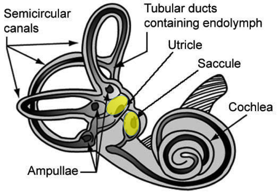
NASA, n.d. The Vestibular System, modified from Wikimedia Commons. Used under a Creative Commons license.
You can see what makes this a complicated structure - all the loops and coils sticking out in all directions! Let's look at the three loops sticking out from the left side, the semicircular canals. These structures detect rotational movement of your head.
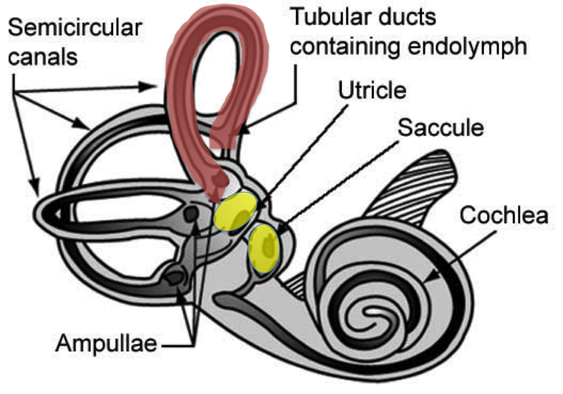
NASA, n.d. The Vestibular System, modified from Wikimedia Commons. Used under a Creative Commons license.
Each semicircular canal is a tube containing fluid. Let's look at the anterior semicircular canal at the top of the picture, highlighted in red. This canal is oriented from the posterior side of the ear to the anterior side, sort of like the handle of a bucket. When you nod your head, you will make the fluid inside this canal move. At the bottom of the canal is an enlarged area called an ampulla, and this is where the hair cells live. They sit down there waiting for the moving fluid to bend their cilia. Like the hair cells in the utricle and saccule, they have a blob of jelly on their cilia - but no otoliths.
You have three semicircular canals in each ear. Each ear has the anterior canal oriented to detect when you nod, the lateral canal oriented horizontally to detect when you turn your head, and the posterior canal oriented left-to-right to detect when you bend your neck to the side.
Between the otolith organs and the semicircular canals, you can always tell where your head is at! If something goes wrong with them, though, you are likely to have poor balance or dizziness (vertigo).
Page 5
When you look at the labyrinth, you can see that the way the chambers are built makes sure that the hair cells in each chamber detect a specific movement. Now we're going to look at how the ear's construction allows a more challenging task - detecting sound waves.

Brocken Inaglory, 2008. Sea Storm in Pacifica. from Wikimedia Commons. Used under a Creative Commons license.
To make sense of this, let's start where the sound is - outside your head. Waves in the air are pounding against your ears.
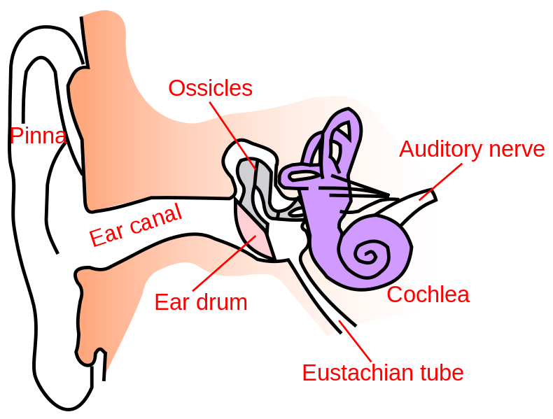
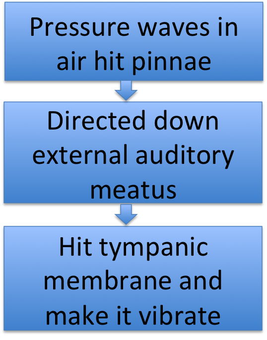
You catch those waves with your external ears, starting with the auricles or pinnae. The bigger they are, the better you can catch the sound waves and funnel them down the ear canal or external auditory meatus to the eardrum or tympanic membrane. Now the waves hit the tympanic membrane, making it vibrate like the head of a drum.
On the other side of the eardrum is a chamber, the middle ear, containing three bones called ossicles. The first of these bones, the hammer or malleus, is attached to the eardrum - so when the eardrum vibrates, so does the malleus. It passes the vibration on to the anvil or incus, which passes it on to the stirrup or stapes, which passes it on to the bony labyrinth where the hair cells live.
Unlike the labyrinth, which is filled with fluid, the middle ear chamber contains air. After all, the eardrum could hardly vibrate if it had fluid pushing on it from behind! Air can enter or leave the middle ear through the eustachian tube, which is connected to your throat. This is important, because you need to keep the air pressure on both sides of the eardrum equal. Otherwise the eardrum might be stretched or even burst.
You experience the eustachian tube when you have to clear your ears after going up in a plane or a high elevator. When you chew gum or otherwise move your jaws, you stretch the tube open and let air pressure equalize, making the pain in your eardrum go away. The down side of the eustachian tube, though, is that it can let bacteria and viruses move from your throat into the middle ear.
Page 6
We've followed the sound waves in the air through the outer ear to the eardrum, and through the middle ear until the stapes transmitted those vibrations to the vestibule. But how does your ear get the vibrations to the right set of hair cells?
Just as with the cells that detect motion, the structure of the inner ear makes this happen.
Let's look back at the inner ear. The hair cells that detect sound live in their own part of the inner ear, the cochlea. And the stapes attaches to the base of the cochlea - so vibrations from the stapes go to those cells only.
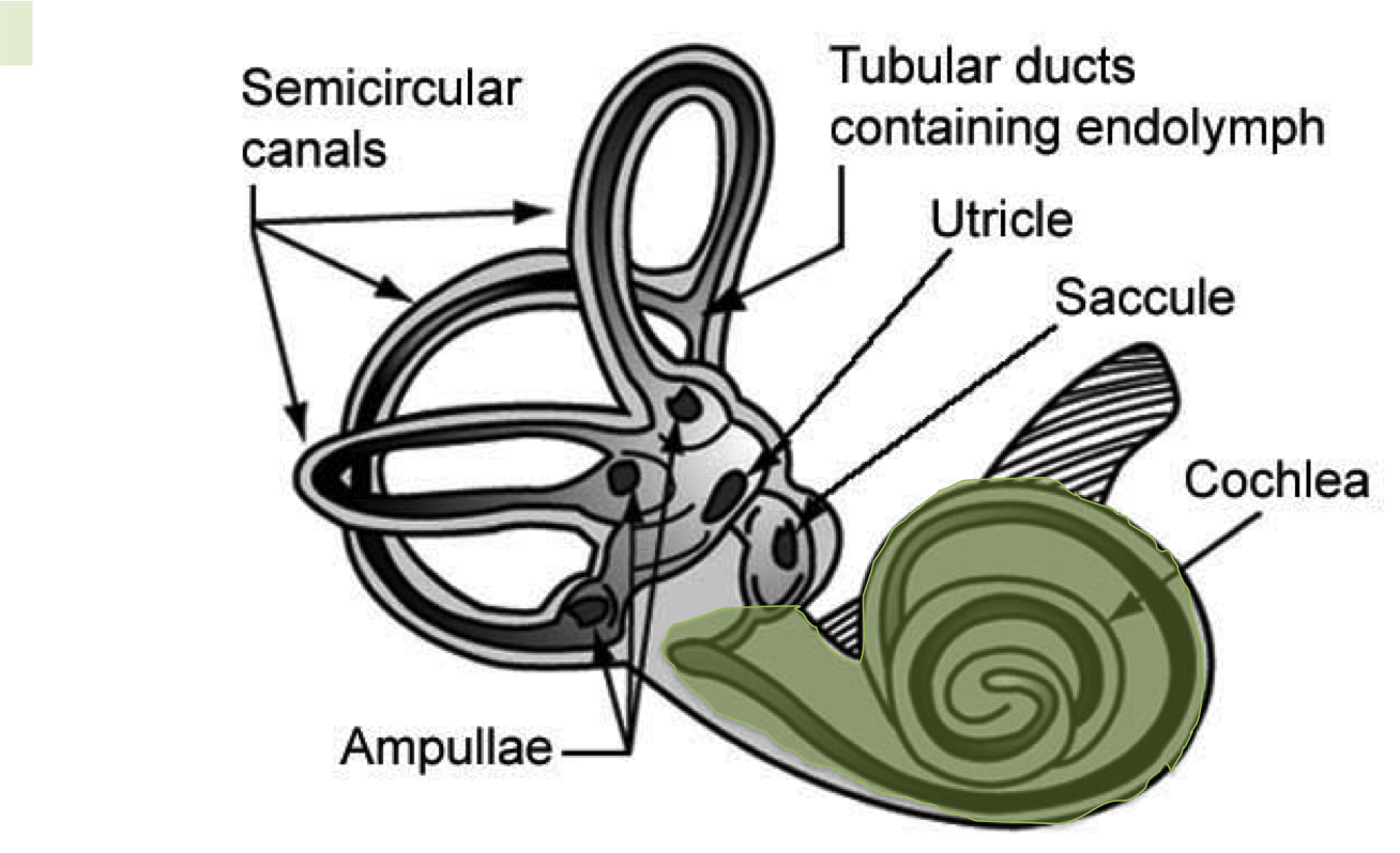
NASA, n.d. The Vestibular System, modified from Wikimedia Commons. Used under a Creative Commons license.
Looking at the picture you can see that the cochlea, like the semicircular canals, is a fluid-filled tube. The auditory hair cells live inside that tube. Like all the other hair cells, their cilia are embedded in something, the tectorial membrane that sticks in from the side of the cochlear duct, like a long shelf. If we unwound the cochlea, then, we would see a long tube running down the center, with hair cells inside it and the tectorial membrane connecting their cilia. This tube is called the cochlear duct.
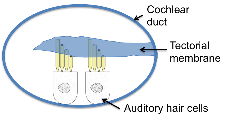
In this cross-section, you can see the hair cells along the lower side of the duct, with their cilia extending upwards into the tectorial membrane.
But how do the vibrations from sound make these hair cells fire? There has to be more to hearing than just hair cells firing, because we can distinguish different sounds. The answer is in the rest of the cochlea, the part outside the cochlear duct.
Page 7
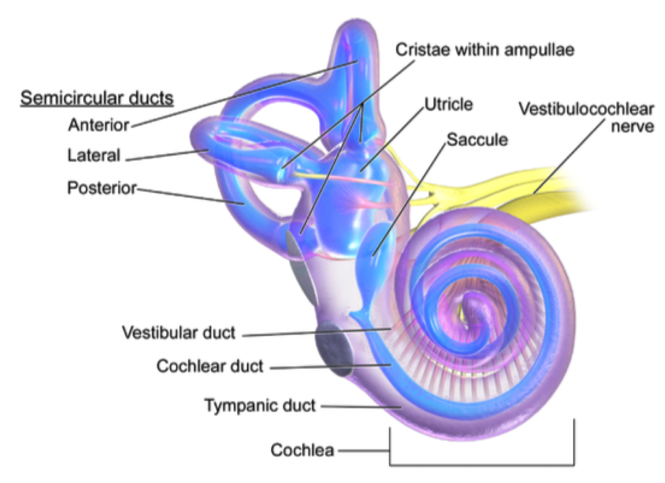
Blausen.com staff. "Blausen gallery 2014". The Internal Ear. Wikiversity Journal of Medicine. Modified from Wikimedia Commons. Used under a Creative Commons License.
In this picture, you can see a little more clearly; there is a space on each side of the cochlear duct. These spaces are actually also tubes full of fluid. In fact, they are one tube bent back on itself. If we uncoiled the cochlea, it would look like this:

You can see the cochlear duct straightened out, running down the center, and the other tube running beside it to its end, turning around, and running back up the other side. On one side, the outer tube is called the tympanic duct, and on the other side it is called the vestibular duct. The cochlear duct and the hair cells living inside it are cushioned on both sides by this tube full of fluid. And it is that outer tube that the stapes attaches to - not the cochlear duct!
Now we have the structure, how does it work?

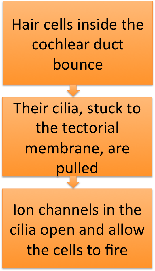
The vibrating stapes pushes on the end of the vestibular duct, making waves in the fluid. Those waves bounce the hair cells in the cochlear duct, so their cilia are pulled and they fire.
Which hair cells will fire? It depends on how far the waves travel down the duct. High-frequency waves (high pitches) don't travel very far, so they make the hair cells near the stapes respond. Low-frequency waves (low pitches) travel further, and stimulate the cells at the far end of the cochlea. This is how you distinguish what notes you have heard - by which auditory hair cells send a message to the brain.
Page 8
Time for a review. Choose the right words to fill in the blanks in this flow chart of sound's passage through the outer ear.
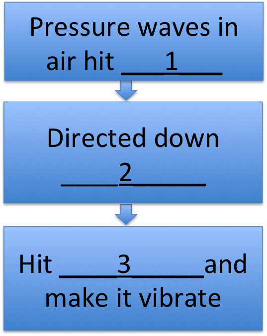
Pressure waves in the air hit the (1): external ears / pinnae / hair cells
The waves are directed down the (2): external auditory meatus / cochlea / eustachian tube
Until they hit the (3): saccule / tympanic membrane / incus
Page 9
Good work! Now choose the right words to fill in the blanks in this part of the flow chart.
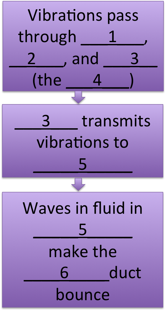
Vibrations from the tympanic membrane are passed to the (1): incus / malleus / hair cells
Which passes them to the (2): incus / cochlea / eustachian tube
Which passes them to the (3): saccule / stapes / incus
All together, these three structures are called the (4): ossicles / otoliths / tympani
The last of these transmits vibrations to the (5) in the inner ear: tympanic duct / vestibular duct / cochlear duct
Waves in the fluid inside this structure cause the (6) contaning the auditory hair cells to bounce: tympanic duct / vestibular duct / cochlear duct
Page 10
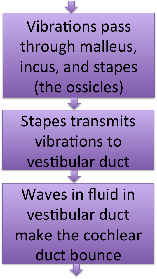
Good work! Now choose the right words to fill in the blanks in the last part of the flow chart.
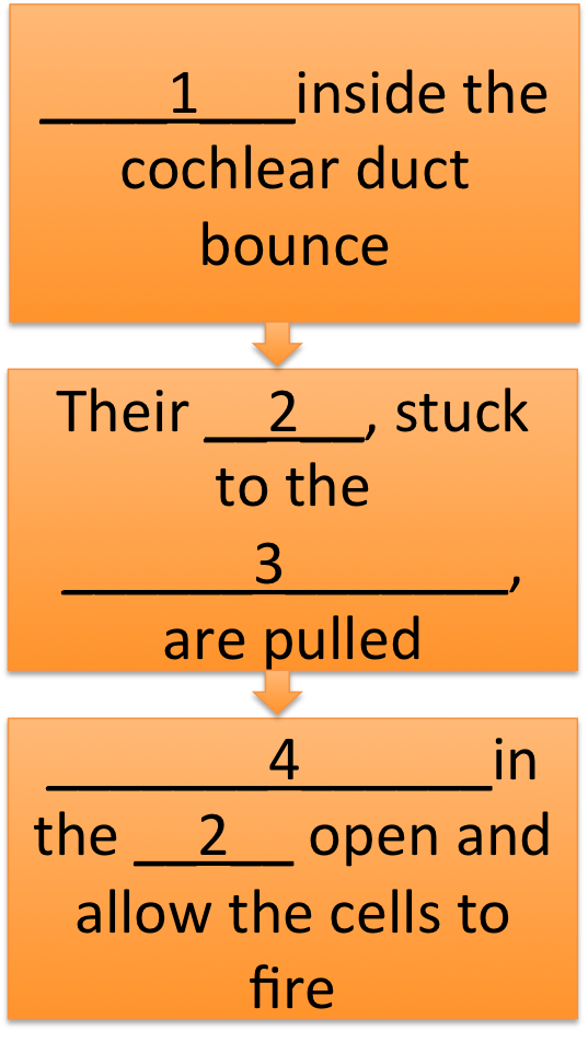
When the cochlear duct bounces, so do the (1) inside it: otoliths / hair cells / eustachian tube
The ends of their (2) are embedded in the (3), so the bouncing pulls them. 2 should be: hair follicle / cochlea / cilia
3 should be: bony labyrinth / utricle / tectorial membrane
This causes (4) to open, allowing the cells to fire and send a message to the brain: ion channels / otoliths / stapes
Page 11
Good work building the flow chart of how hearing works!

Can you create a similar chart to explain how your inner ears detect head motion? Try this one:
Someone asks you a question just as you get on the elevator. You shake your head 'no' in reply. Then the door closes and you realize the elevator is going down instead of up. You tip your head back to look up at the floor numbers. How did your brain find out about all these movements? Here's a diagram to remind you of the names of structures in the labyrinth.

Blausen.com staff. "Blausen gallery 2014". The Internal Ear. Wikiversity Journal of Medicine. Modified from Wikimedia Commons. Used under a Creative Commons License.
Try to create a flow chart right now, before looking at the answer on the next page.
Page 12

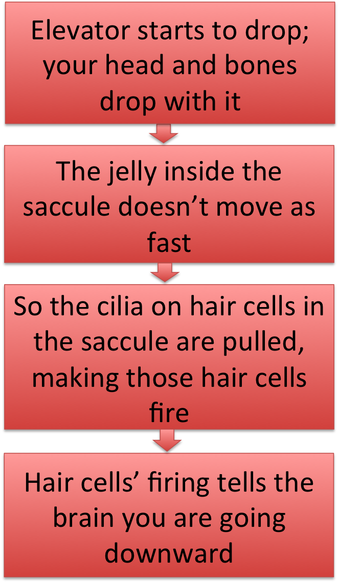
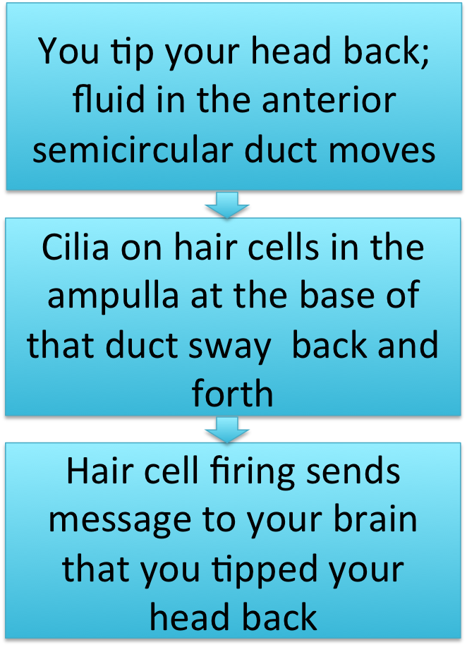
Notice there's a lot in common in these three charts. In the first and third (when you shake your head and tip it up), what matters is that the fluid in the semicircular canals moves. Shaking your head moves the fluid in the lateral semicircular canal, and tipping it moves fluid in the anterior semicircular canal. In each case, the cells in the ampulla at the bottom of the canal fire.
When the elevator drops, your saccule comes into play; once again, its movement makes the hair cells in it fire.
Can you think of some details that could be added to this flow chart to make it more accurate?
You've finished the tutorial on ears and hearing. As you study for the assessment, ask yourself - what would happen to a person if one of these structures stopped working properly?
This tutorial is based on material from Fox, S.I., 2013. Human Physiology, 13th ed. McGraw-Hill.
