This tutorial covers how the spleen is involved in the immune response.
You can navigate through this tutorial using the buttons at the top of the screen.
The tutorial will ask you questions. Click on your chosen answer to see feedback; click the answer again to make the feedback disappear. When you're finished with one page, click the navigation button for the next page to move ahead. If a word is bolded; click on the word to see additional information; click the word again to make this information disappear.
Have fun! Click on button '1' to see the first page of the tutorial.
Page 7
Up to this point you have been filling in the blanks to the immune response pathway. Test your knowledge of the immune response by filling in the blank in this review pathway.
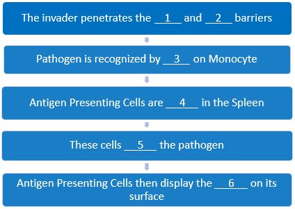
4 should be activated/phagocytosed/presented
5 should be engulf/digest/phagocytize
6 should be epitope/antibody/antigen
Page 8
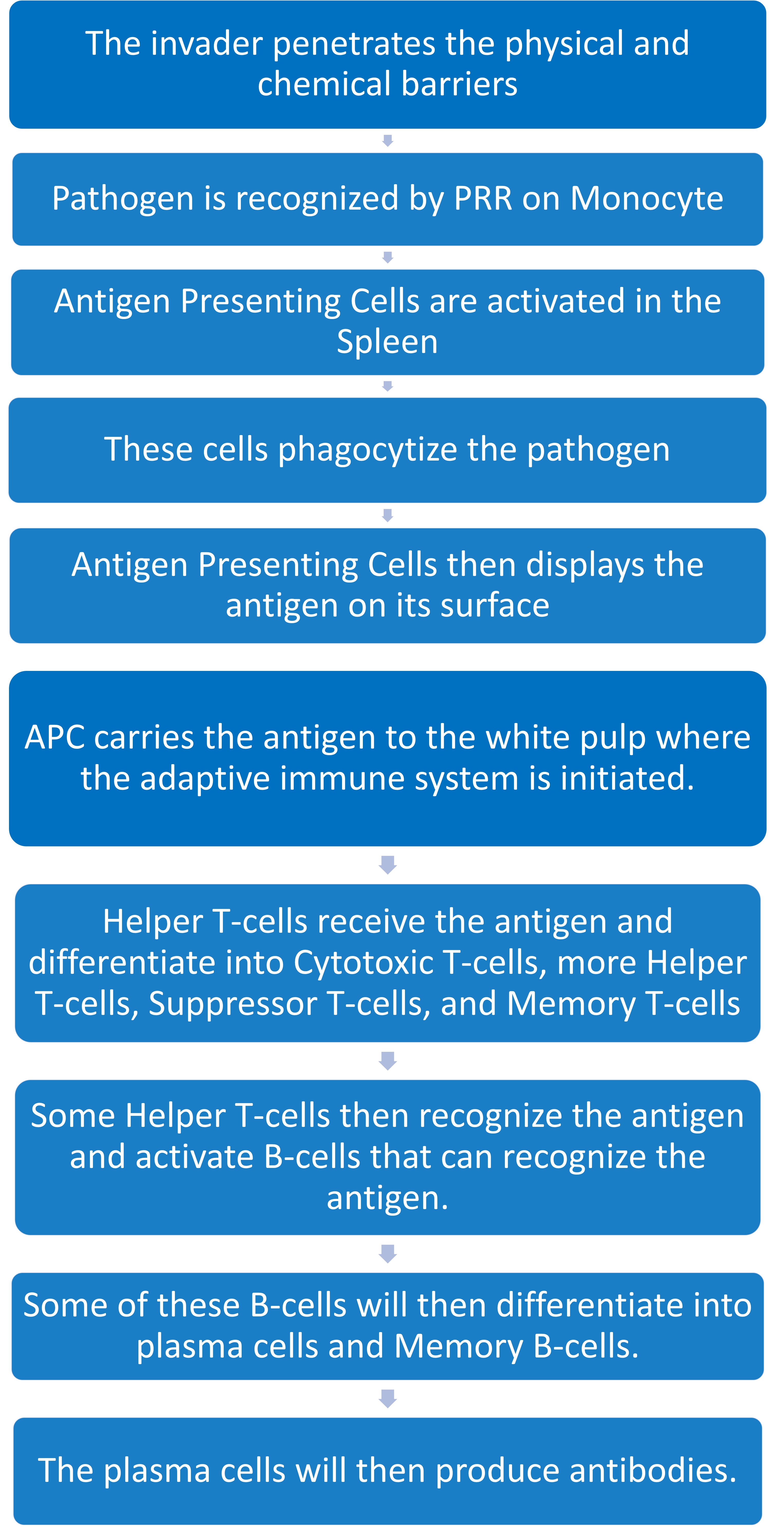
Immune response pathway continued. You should be able to fill in the blanks of the adaptive immune response. For a review of the adaptive immune response click here
.jpg)
7 should be white pulp/red pulp/lymph node
8 should be destroyed/located/ initiated
9 should be Helper T-cells/Memory T-cells/Cytotoxic T-cells
10 should be Cytotoxic T-cells/B-cells/APCs
11 should be Cytotoxic T-cells/APCs/Suppressor T-cells
12 should be Monocytes/Memory T-cells/APCs
13 should be B-cells/plasma cells/APCs
14 should be Helper B-cells/plasma cells/more B-cells
15 should be Memory B-cells/APCs/antibodies
16 should be cytokines/antigens /antibodies
Page 9
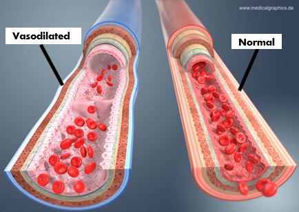
Wikimedia Commons (n.d.). Red Pulp and White Pulp of the Spleen. Retrieved May 17, 2019, from http://www.medicalgraphics.de/en/free-pictures/organs/comparison-blood-vessels-arteries-veins-dark-429.html
APC also induce the inflammatory response by releasing cytokines which signal for additional leukocytes to come to the site of infection. These leukocytes enter from blood vessels and once in the spleen they fight the infection via phagocytosis/other mechanisms. Each time they kill a pathogen or cells are damaged more proinflammatory cytokines are released.
Proinflammatory cytokines also causes vasodilation which increases / decreases / doesn't affect blood flow and increases / decreases / doesn't affect permeability; allowing the cells to move to the spleen faster.
Page 10
Mary Jane went to the doctor and was told she had an enlarged spleen.
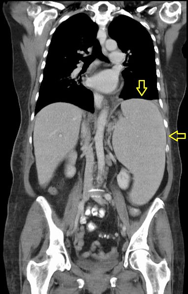
Wikimedia Commons. (n.d.). Splenomegalie bei CLL (labeled). Retrieved May 17, 2019, from https://commons.wikimedia.org/wiki/File:Splenomegalie_bei_CLL_(labeled).jpg
Why did Hepatitis B cause her spleen to be enlarged?
A. Her spleen had proliferating immune cells and cells being called in to fight the infection
Page 11
The doctor prescribed Mary Jane medication that helped fight the infection. Her liver was able to recover, and spleen is back to normal size. Mary Jane lives to see another day!
Page 1
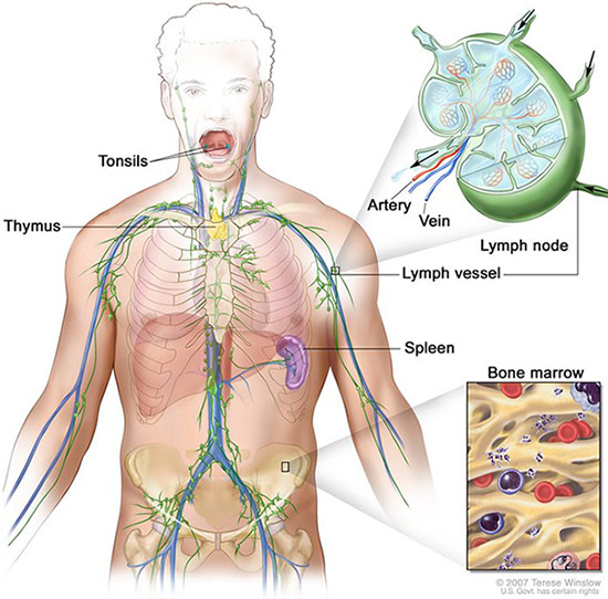
Lymphatic System by U.S. National Cancer Institute's SEER Program, Public Domain.
Hepatitis B is a dangerous blood borne pathogen that is contracted through blood, semen, and other bodily fluids. It is caused by a virus that mainly affects the liver but is known to cause splenomegaly (an enlarged spleen). In this case study you will take a closer look at the role of the spleen in the immune response.
As a reminder, when pathogens enter the body (unless they are in the blood) the pathogen will be processed through the lymphatic system. As shown in green, the lymphatic system consists of various parts of the body,(thymus, bone marrow, lymph nodes, etc) that work together to detect pathogens. This case study begins examining the patient, Mary Jane, and the Hepatitis B pathogen currently in her circulatory system.

The first main protection our bodies provides from invaders are our skin and mucous membranes which are known as physical barriers. At these surfaces our bodies uses chemical barriers such as sweat, saliva enzymes, and antimicrobial proteins to prevent pathogens from entering the body.
Page 2
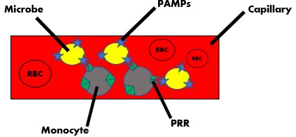
The roles of the spleen are to repair/destroy damaged RBC and scan the blood for pathogens. The RBCs are processed in the red pulp of the spleen, while the white pulp processes the pathogen to initiate the adaptive immune response. In both these areas, WBC use special cell surface receptors called Pathogen Recognition Receptors (PRRs) to identify generic Pathogen Associated Molecular Patterns (PAMPs).
Specifically, PAMPs are located on the surface of microbes and are recognized by immune cells via their unique patterns - referring to their shapes and chemistry. PRR are located extracellularly and intracellularly on all nucleated cells. These bind to ligands, (PAMPs) on microbes. One of the main types of WBCs in blood are Monocytes. They use their PRR to look for surface markers on microbes inside the capillary. Once found, the Monocyte will bind to the microbe and carry it away.
 3 should be
Page 3
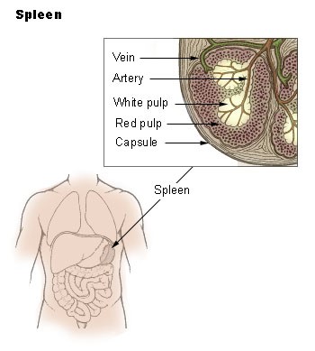
National Cancer Institute SEER training modules (n.d.) Spleen. Public domain image.
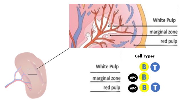
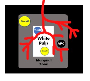
The monocyte with the attached microbe is now circulating to the spleen, the spleen is divided into three sections: the red pulp, the marginal zone, and the white pulp. The spleen is only connected to the circulatory system not the lymphatic system. Thus, the only way in is through the splenic artery where the monocytes will be dumped with the blood into the marginal zone. Located here are macrophages (APC) and B-cells.
All RBCs will be sent to the red pulp that has a sinusoid network with macrophages and lymphocytes (T-cells, B-cells, and NK cells). There the RBCs will either be repaired or destroyed. For a review of Red Blood Cell Life Cycle, click here The blood will then exit through the splenic vein back into the circulatory system.
Modified from: Figure 2f from: Irimia R, Gottschling M (2016) Taxonomic revision of Rochefortia Sw. (Ehretiaceae, Boraginales). Biodiversity Data Journal 4: e7720. https://doi.org/10.3897/BDJ.4.e7720
Anything that is not a RBC it will be taken to the white pulp which will be discussed in slide six. To see what APCs do with the antigen continue with slide four and five.
Page 4
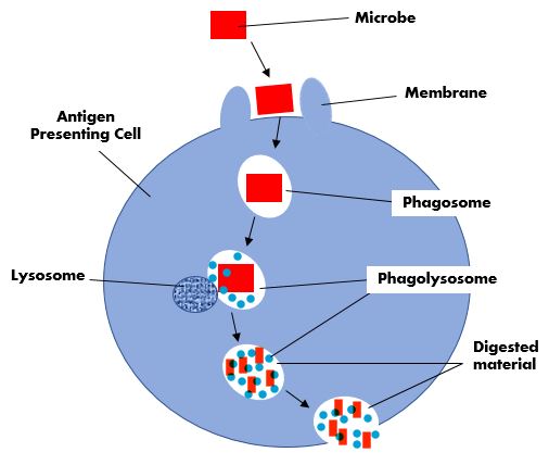
In the marginal zone, the monocyte passes the microbe off by sending cytokines to the Professional Antigen Presenting Cells, (APCs) who begin phagocytizing the pathogen. Cytokines are signals released by cells to communicate to other cells. These signals are actually molecules received via cytokine receptors.
The process of phagocytosis begins with the APC's membrane extending around the microbe. The ingestion of the microbe by the phagocyte results in the formation of a phagosome. The phagosome is then fused with lysosome to form a phagolysosome. Inside the phagolysome the microbe is degraded by ROS which stands for Reactive Oxygen Species. This waste material is then expelled out of the APC.


Page 5
Phagocytosis Review: In the chart below, select the answer that best completes the sentence.
The APC's cell wall / membrane / phagosome extends around the phagosome / membrane / microbe
The phagosome / membrane / microbe is internalized and a phagosome/lysosome/phagolysosome is created.
The phagosome/lysosome/phagolysosome is filled with phagosome/lysosome/phagolysosome resulting in a phagosome/lysosome/phagolysosome.
This causes a release of reactive oxygen/nitrogen species that creates / inhibits / destroys the phagosome / membrane / microbe
The microbe / digested material / ROS is then released.
Page 6
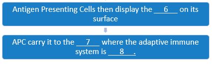
6 should be epitope/antibody/antigen
7 should be white pulp/red pulp/lymph node8 should be destroyed/located/ initiated
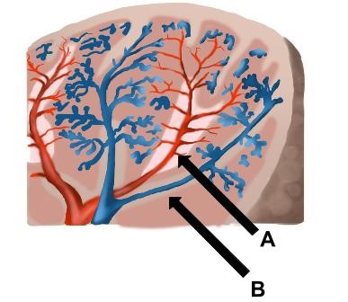
Oláh, I., & Glick, B. (1982). Splenic white pulp and associated vascular channels in chicken spleen. American Journal of Anatomy, 165(4), 445-480. doi:10.1002/aja.1001650408
white pulp/red pulp/marginal zone
In the spleen, B is pointing to?
white pulp/red pulp/marginal zone
Which type of cell is located in all portions of the spleen? (Hint: On slide 3 there was a picture with all the types listed out)
T-cells/B-cells/Antigen Presenting Cells